MRI CD
Today I found out again that certain people love me, and still others who don't know me, do care. Thanks, everybody.
The neurologist shared with us the impressions report that the radiologist doing my MRI had. This was not the same person who requested the study; she is on vacation at the moment. Basically they say that the unusual brightness they saw on the first MRI is less bright now.
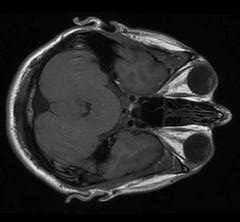
Hyperintense signal in the posterior aspect of the internal capsules bilaterally right greater than left, extending inferiorly to the level of the cerebral peduncles and superiorly to the cortex and the parietal centrum semiovale. The pattern of distribution is identical to the previous examination, however, the findings are more subtle in signal characteristics than on the previous examination.
I never knew I had peduncles. I wonder if women have them?
Back when I had a bright signal they said it didn't mean anything bad. And now that the signal is less bright they say it doesn't mean anything good. Bright, or not bright, why should I care? But they want me to feel encouraged.
I may have messed up by declining the contrast agent. I did so because, after the first MRI, I was told that the contrast agent had not made any difference in the scan results. Predictably, though, it turns out that they do additional scans from a certain angle when using the contrast agent. And this time they did not do those scans, which my neurologist and the radiologist writing the paper want to compare to last year's scans. I'm willing to go in for another MRI if she asks for it, since I was able to tolerate the procedure pretty well this most recent time.
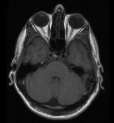
I offer the second photograph in this post because I believe it shows the white stuff in those two narrow streaks towards the center of the brain. The more intense white stuff all around the perimeter of the brain is presumably completely normal and healthy. Whatever. I didn't ask. They would have told me.
I believe that this scan is one that shows that the effect is greater on my right than on my left. Keep in mind that the scan is displayed from the perspective of the feet, looking up to the top of the skull. So apparent right and left are reversed. The scan shows more damage on the right side of my brain, hence the left side of my body (controlled by the right side of my brain) is more messed up. Thing is, though, in most people with ALS, the scan does not show any special signal along the motor pathway. So the whole thing seems pointless to me. But then, great science usually comes from studying things thought not to matter.
Oh, by the way, I have decided to stop taking ginseng because, very roughly speaking, the period of my latest stepwise decline coincides with my adding ginseng back into my pill regimen.
The neurologist had a puppy in the office that day. We played with it.
The good news is, I still don't have brain cancer!
posted by brainhell at 5:39 PM
![]()

<< Home